Health
When Your Doctor Recommends Imaging of Your Pelvic Floor

Health Points
- Defecography uses X-ray or MRI imaging to diagnose pelvic floor disorders affecting bowel function
- The test helps identify structural problems like prolapse or rectocele that may cause chronic constipation
- While the procedure may feel uncomfortable, it provides critical information for treatment planning
For adults experiencing persistent bowel difficulties, doctors sometimes recommend a specialized imaging test that examines how the pelvic floor muscles and organs function during bowel movements. This diagnostic procedure, called defecography, has become an important tool for identifying structural problems that traditional exams might miss.
Defecography creates detailed images of the rectum, pelvic floor, and surrounding structures while a patient attempts to evacuate contrast material from the rectum. The test can be performed using either X-ray technology (fluoroscopic defecography) or magnetic resonance imaging (MRI defecography), with each method offering distinct advantages for different clinical situations.
“This is really the gold standard for understanding what’s happening mechanically when someone has difficulty with bowel movements,” explains Dr. Sarah Mitchell, a colorectal surgeon at Johns Hopkins Medicine. “We can see in real time whether the pelvic floor is relaxing properly, if there’s a structural prolapse, or if the rectum is emptying completely.”
The test typically becomes necessary when patients experience chronic constipation that hasn’t responded to dietary changes or medication, a persistent sensation of incomplete evacuation, or symptoms suggesting pelvic organ prolapse. Women over 50 who have had multiple vaginal deliveries face higher risk for pelvic floor disorders that might warrant this type of imaging.
During fluoroscopic defecography, patients receive a thick paste containing barium—a contrast material visible on X-rays—inserted into the rectum. The radiologist then takes continuous X-ray images while the patient sits on a specialized commode and attempts to evacuate the contrast material. The procedure typically takes 15 to 30 minutes and exposes patients to a small amount of radiation comparable to other routine X-ray examinations.
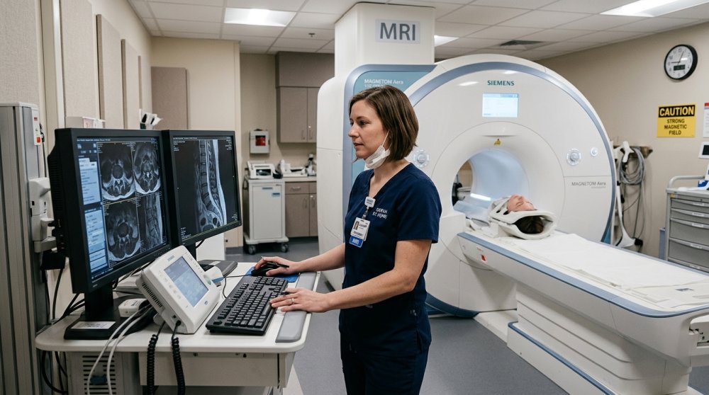
MRI defecography follows a similar process but uses magnetic imaging instead of X-rays. Patients receive a gel contrast material and lie on their side within the MRI scanner. This approach provides more detailed images of soft tissues and avoids radiation exposure, though it takes longer to complete and costs more than the X-ray version.
“The MRI gives us exceptional detail of the pelvic floor muscles, ligaments, and all three compartments—bladder, uterus or vagina, and rectum,” notes Dr. Mitchell. “For complex cases or younger patients where radiation is a concern, it’s often worth the additional time and expense.”
Both versions of the test can identify several conditions affecting bowel function. Rectocele, where the rectal wall bulges into the vagina, commonly appears in women who’ve experienced childbirth trauma to the pelvic floor. Rectal prolapse, where the rectum telescopes down through the anus, can cause bleeding and difficulty controlling bowel movements. The imaging may also reveal excessive straining of the pelvic floor muscles that prevents proper evacuation.
Preparation for defecography typically involves following a clear liquid diet the day before and using enemas to empty the bowels. Patients should inform their doctor about any medications they take, particularly blood thinners, and women of childbearing age should confirm they’re not pregnant before X-ray defecography.
While many patients feel embarrassed about this test, medical professionals emphasize that it provides information unavailable through other means. Standard colonoscopy or physical examination can’t assess how the pelvic floor functions during attempted bowel movements, making defecography uniquely valuable for certain diagnoses.
“I always tell patients this is a medical test like any other,” says Dr. Mitchell. “The technologists and physicians who perform these studies do them every day. There’s no reason to feel self-conscious about a test that could dramatically improve your quality of life.”
Recovery from the procedure itself is immediate, though patients may experience some rectal fullness or mild cramping as the contrast material passes. Drinking plenty of water helps the body eliminate the barium more quickly. Results typically become available within a few days, when the radiologist completes their detailed analysis of the images.
The findings from defecography guide treatment decisions ranging from pelvic floor physical therapy to surgical repair of prolapsed organs. Many patients who’ve struggled with bowel symptoms for years finally receive accurate diagnoses that lead to effective treatment plans. Conservative approaches like biofeedback therapy work well for some conditions, while others may require surgical intervention for optimal results.
For adults over 40 experiencing persistent bowel difficulties despite trying standard treatments, defecography offers valuable diagnostic clarity. While the test may feel awkward, the detailed functional information it provides helps physicians develop targeted treatment strategies that address the root cause of symptoms rather than just managing discomfort.
Understanding what happens during the procedure and why doctors recommend it can reduce anxiety and help patients feel more comfortable with this important diagnostic tool. When chronic symptoms significantly impact daily life, accurate diagnosis becomes the essential first step toward lasting improvement.
