Health
Why Doctors Are Turning to This Advanced Imaging Technology for Earlier Lung Cancer Detection
Health Points
- PET scans combine metabolic and anatomical imaging to detect lung cancer earlier and more accurately than traditional CT scans alone
- The procedure uses a radioactive tracer that highlights cancer cells, helping doctors distinguish between benign and malignant growths
- PET scanning plays a critical role in staging cancer, planning treatment, and monitoring how well therapy is working
When it comes to detecting lung cancer, timing can make all the difference. Doctors now have access to sophisticated imaging technology that goes beyond traditional X-rays and CT scans—and it’s changing how early-stage lung cancer is found and treated.
Positron emission tomography, commonly called a PET scan, offers a unique window into the body’s cellular activity. Unlike standard imaging that simply shows anatomical structures, PET scans reveal how tissues are functioning at a metabolic level, making them especially valuable for identifying cancerous growths.
The technology works by introducing a small amount of radioactive tracer—typically a form of glucose—into the bloodstream. Cancer cells, which metabolize sugar at higher rates than normal cells, light up on the scan, creating a visual map that helps physicians pinpoint problem areas with remarkable precision.
For patients over 40, understanding this diagnostic tool becomes increasingly relevant. Lung cancer remains one of the most common cancers in the United States, and early detection significantly improves treatment outcomes and survival rates.
How the Scanning Process Works
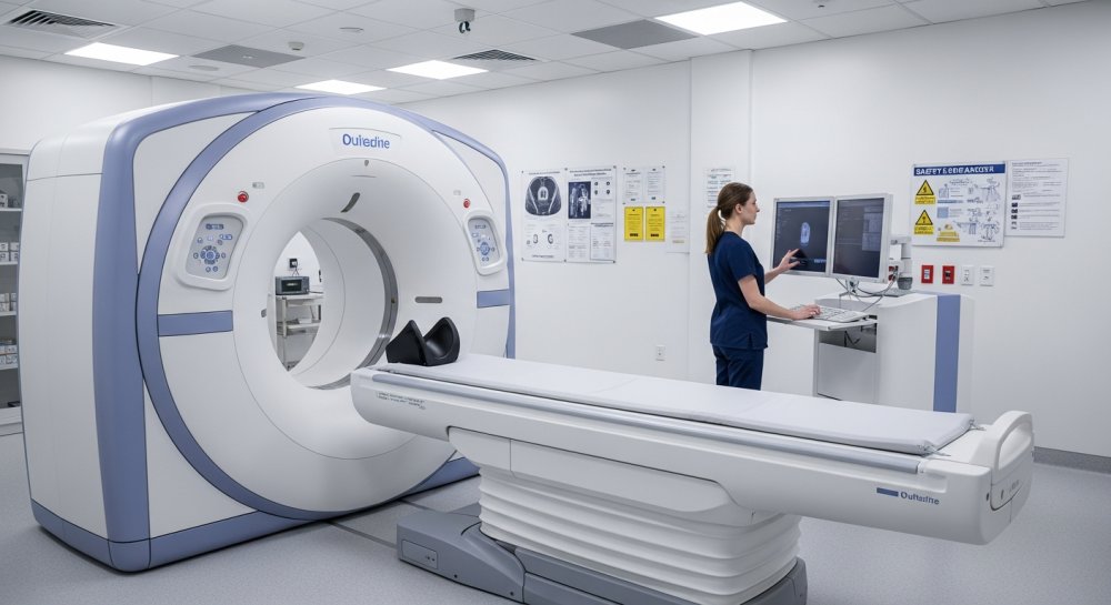
Before undergoing a PET scan, patients receive an injection of the radioactive tracer and then rest quietly for about an hour while the substance circulates through the body. The actual scanning takes approximately 30 to 45 minutes, during which patients lie still on a table that moves slowly through a large, doughnut-shaped machine.
The procedure itself is painless and non-invasive. Patients don’t experience any sensations from the radioactive tracer, which contains only a tiny amount of radiation—roughly equivalent to what you’d receive from two standard chest X-rays.
Many medical centers now use combined PET-CT scanners, which merge metabolic information from the PET scan with detailed anatomical images from a CT scan. This combination provides doctors with both the location and the biological behavior of suspicious areas in a single examination.
When Doctors Recommend PET Scanning
Physicians typically order PET scans when other imaging tests have revealed a suspicious lung nodule or mass. The scan helps determine whether the abnormality is likely cancerous or benign, potentially sparing patients from unnecessary biopsies or surgeries.
The technology proves especially valuable for cancer staging—determining how far the disease has spread. PET scans can identify cancer that has traveled to lymph nodes or distant organs, information that’s crucial for developing an effective treatment plan.
For patients already diagnosed with lung cancer, PET scans serve multiple purposes throughout the treatment journey. Doctors use them to plan radiation therapy with precision, evaluate how well chemotherapy or other treatments are working, and monitor for cancer recurrence after initial treatment succeeds.
Understanding the Limitations
While PET scans offer powerful diagnostic capabilities, they’re not perfect. Some non-cancerous conditions can cause increased metabolic activity that mimics cancer on a scan, including infections, inflammation, and recent injuries.
Very small tumors—typically those smaller than 8 millimeters—may not absorb enough tracer to appear on a PET scan. In these cases, doctors rely on other imaging techniques and clinical judgment to determine the best course of action.
The test also requires specific patient preparation. People must fast for several hours before the scan because eating raises blood sugar levels, which can interfere with how the radioactive tracer distributes throughout the body and potentially affect image quality.
What the Results Mean
After the scan, a radiologist trained in nuclear medicine analyzes the images and sends a detailed report to the ordering physician. Areas of high tracer uptake appear as bright spots on the scan, indicating increased metabolic activity that could signal cancer.
However, a positive PET scan doesn’t automatically confirm cancer. Doctors consider PET results alongside other test findings, medical history, and sometimes tissue samples obtained through biopsy before making a definitive diagnosis.
For many patients, PET scanning provides reassurance when results come back negative. A scan showing no abnormal uptake patterns in a suspicious lung nodule often means the growth is benign, allowing patients and doctors to pursue less aggressive monitoring approaches.
The Role in Modern Lung Cancer Care
As medical technology advances, PET scanning continues to evolve. Newer tracers beyond the standard glucose-based compound are being developed to target specific types of cancer cells with even greater accuracy.
The integration of artificial intelligence and machine learning is also enhancing how doctors interpret PET scan results. These tools help identify subtle patterns that might escape the human eye, potentially catching cancers at even earlier stages.
For individuals at high risk for lung cancer—including longtime smokers, those with significant secondhand smoke exposure, or people with a family history of the disease—understanding available diagnostic tools empowers more informed conversations with healthcare providers about screening and early detection strategies.
While the thought of undergoing cancer testing can feel overwhelming, modern imaging technologies like PET scanning represent significant progress in the fight against lung cancer. They offer hope through earlier detection, more precise treatment planning, and better monitoring of how well therapies work—all factors that contribute to improved outcomes and quality of life for patients facing this challenging diagnosis.