Health
The Eye Test That Reveals Macular Degeneration Before You Notice Symptoms

Health Points
- Regular comprehensive eye exams can detect macular degeneration years before vision loss begins
- Multiple specialized tests work together to diagnose the condition and determine appropriate treatment
- Early detection significantly improves the chances of preserving central vision
Macular degeneration affects millions of Americans, particularly those over 50, yet many people don’t realize they have the condition until significant vision changes occur. The good news is that eye care professionals have several powerful diagnostic tools that can identify the disease in its earliest stages—often before any noticeable symptoms appear.
The macula, a small area in the center of the retina, is responsible for sharp, detailed central vision used for reading, driving, and recognizing faces. When this tissue begins to deteriorate, it can lead to progressive vision loss.
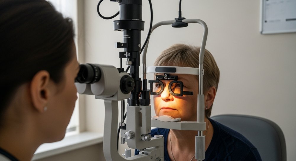
A comprehensive dilated eye examination forms the foundation of macular degeneration diagnosis. During this exam, an ophthalmologist or optometrist uses special drops to widen the pupils, allowing a clear view of the retina and macula. This simple procedure enables doctors to spot early warning signs like drusen—yellow deposits under the retina that indicate age-related macular degeneration (AMD).
The Amsler grid test provides a quick screening tool that patients can even use at home. This simple chart consists of a grid pattern with a central dot. When looking at the grid, people with macular degeneration may notice wavy lines, blank spots, or distorted areas—signs that warrant immediate professional evaluation.
For more detailed imaging, doctors turn to optical coherence tomography (OCT). This non-invasive technology uses light waves to capture high-resolution, cross-sectional images of the retina. OCT can reveal fluid accumulation, retinal thickness changes, and structural abnormalities invisible to the naked eye, making it invaluable for both diagnosis and monitoring disease progression.
Fluorescein angiography offers another diagnostic avenue, particularly for detecting wet AMD, the more aggressive form of the condition. In this procedure, a special dye is injected into an arm vein, and as it travels through the blood vessels in the eye, a camera captures detailed images. These photos reveal abnormal blood vessel growth and leakage—hallmarks of wet macular degeneration that require prompt treatment.
Some practices also employ fundus autofluorescence imaging, which measures natural fluorescent properties of the retina without requiring dye injection. This technique helps identify areas of retinal stress or damage and can detect changes associated with early AMD.
Genetic testing has emerged as an additional tool in assessing macular degeneration risk. Certain genetic variants significantly increase the likelihood of developing AMD, and knowing one’s genetic profile can inform screening frequency and lifestyle modifications.
The American Academy of Ophthalmology recommends comprehensive eye exams every one to two years for adults over 65, and more frequently for those with risk factors including family history, smoking, obesity, and cardiovascular disease. Earlier detection means more treatment options and better outcomes.
Vision changes don’t happen overnight, and neither does macular degeneration. But with regular screening and the advanced diagnostic tools available today, eye care professionals can catch this condition early—when intervention can make the most difference in preserving the clear, central vision that enriches daily life.
