Health
The Vision Condition That Starts in Childhood and Can Lead to Blindness
Health Points
- Myopic macular degeneration occurs when severe nearsightedness causes permanent damage to the retina’s central portion
- The condition typically begins with childhood myopia that worsens over time, affecting about 3% of the global population
- Early detection through regular eye exams and specialized imaging can help slow progression and preserve vision
For millions of Americans living with severe nearsightedness, the challenge goes far beyond remembering where they put their glasses. When myopia reaches extreme levels, it can trigger a serious condition called myopic macular degeneration—a form of vision loss that can permanently alter lives.
This degenerative eye disease develops when the eyeball grows too long during childhood and adolescence, stretching and thinning the retina at the back of the eye. Over time, this stretching damages the macula, the small central area responsible for sharp, detailed vision we use for reading, recognizing faces, and driving.
Unlike the age-related macular degeneration that affects seniors, myopic macular degeneration strikes earlier in life. It typically begins with high myopia in childhood that progressively worsens.
The condition affects approximately 3% of people worldwide, though rates vary significantly by region. Asian populations face the highest risk, with some studies showing prevalence rates as high as 10% in certain areas.
Eye care professionals emphasize that not everyone with nearsightedness will develop this condition. It primarily threatens those with severe myopia—generally a prescription of -6.00 diopters or stronger.
“The eyeball essentially grows too much during development,” explains the medical community’s understanding of the condition. “This excessive elongation stretches all the layers of the retina, making them thinner and more vulnerable to damage.”
As the retina stretches, several complications can arise. Abnormal blood vessels may grow beneath the retina, leading to bleeding and scarring. The stretched tissue becomes more prone to tears and detachment. In advanced cases, permanent blind spots develop in central vision.
Warning signs include sudden increases in floaters, flashes of light, distorted or wavy vision, difficulty seeing in low light, and progressive loss of central vision. These symptoms warrant immediate evaluation by an eye care specialist.
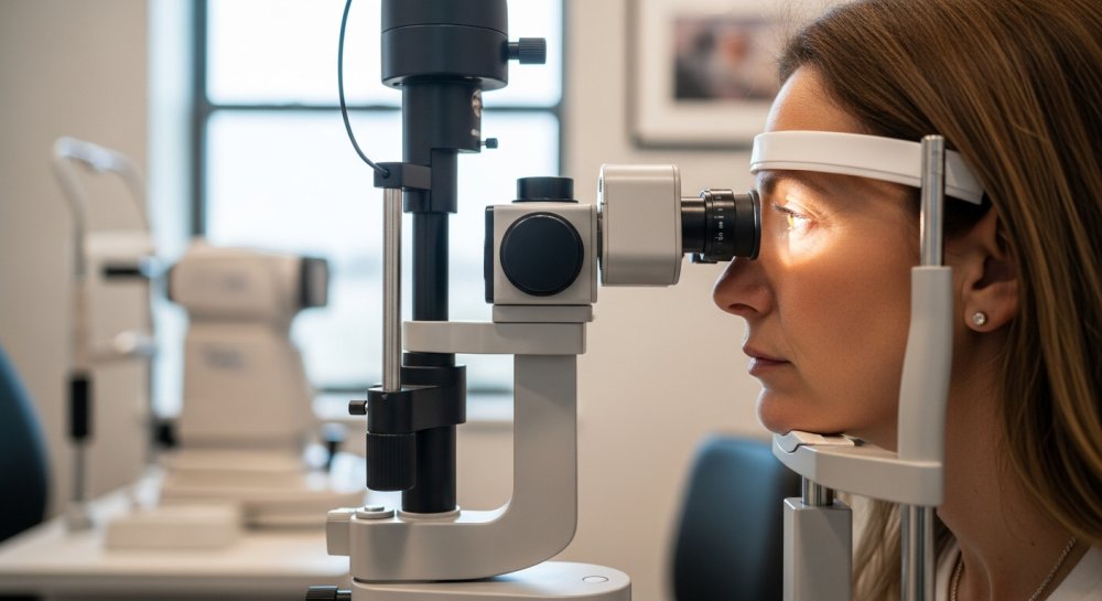
Early detection makes a significant difference in outcomes. Comprehensive eye exams combined with specialized imaging can identify changes before symptoms appear.
Optical coherence tomography creates detailed cross-sectional images of the retina, revealing thinning and structural changes. Fluorescein angiography uses injectable dye to highlight abnormal blood vessel growth. Fundus photography documents changes over time for comparison.
Treatment focuses on slowing progression and managing complications rather than reversing damage already done. For children showing signs of rapidly progressing myopia, specialized contact lenses and low-dose atropine eye drops have shown promise in slowing eyeball elongation.
When abnormal blood vessels develop, anti-VEGF injections can halt their growth and prevent further vision loss. These medications block the protein that promotes new blood vessel formation.
Regular monitoring remains essential, typically requiring eye exams every three to six months for those at high risk. This vigilance allows specialists to intervene quickly when changes occur.
Beyond medical treatment, lifestyle modifications support eye health. Adequate outdoor time during childhood may reduce myopia progression—studies suggest at least two hours daily of natural light exposure. Limiting extended close-up work and taking regular breaks from screens helps reduce eye strain.
The condition carries significant implications for daily life. Those affected may struggle with driving, especially at night. Reading requires stronger magnification or assistive devices. Workplace accommodations become necessary for tasks requiring detailed vision.
For families with a history of severe myopia, early intervention offers the best protection. Children should receive comprehensive eye exams starting at age six months, with follow-ups at three years and before starting school. Those with myopic parents benefit from annual exams throughout childhood and adolescence.
Research into new treatments continues advancing. Scientists are exploring gene therapy approaches and medications that might strengthen retinal tissue against the effects of stretching.
The financial burden of managing this condition extends beyond standard vision correction. Specialized imaging, frequent monitoring appointments, and potential surgical interventions create ongoing expenses that many insurance plans cover only partially.
Support groups and low vision specialists help those affected maintain independence and quality of life. Adaptive technologies—from smartphone apps that enhance contrast to electronic magnifiers—expand possibilities for work and recreation.
Public health experts emphasize the growing urgency of myopia management as rates increase globally. Projected trends suggest that by 2050, nearly half the world’s population could be myopic, with a substantial portion at risk for high myopia and its complications.
Parents play a crucial role in prevention and early detection. Encouraging outdoor play, limiting screen time for young children, ensuring proper lighting for reading and homework, and scheduling regular eye exams can make a lasting difference.
For adults already living with high myopia, staying current with eye exams and immediately reporting any vision changes remains the most effective strategy for preserving sight. While myopic macular degeneration presents serious challenges, advances in detection and treatment continue improving outcomes for those affected.